Results You searched for: Image Cytometry Results Displayed: 1 - 4 of 4
Celígo® Image Cytometry
Every Cell. Every Well.
The bench-top Celigo image cytometry system provides high-throughput whole-well imaging and quantitative data through image analysis in bright field and up to four fluorescent channels, for a wide variety of cell-based assays. It is routinely used to investigate adherent and suspension cells, 3D tumor spheroids and colonies of iPSC and cancer stem cells. It is compatible with microwell plates from 6 to 1536-well and T-flask formats.
The work-flow based intuitive software provides concurrent imaging and analysis; kinetic analysis such as time-lapse growth tracking and flow cytometry-like gating analysis and reporting of cell populations. Cell images of specific populations may be displayed with color overlays through a gating selection.
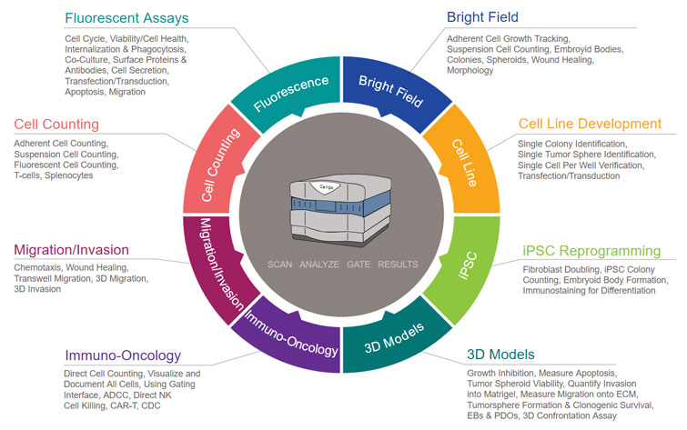
The Celigo product allows users to perform high-speed, fully automated imaging and quantification of a wide range of cell types across complex sample types. It enables an extensive menu of applications including label-free cell counting, confluence-based cell growth tracking, killing assays, apoptosis, cell cycle analysis, migration and invasion assays, as well as cellular assays for receptor internalization, protein expression and detection, phosphorylation and phagocytosis.
Whole-Well, High Resolution Images Acquired at High Speed
Proprietary optical design enables uniform illumination and consistent edge contrast
Image and count every cell in each well: 0 - 100,000 cells/96-well
5 imaging channels with bright field and 4 fluorescent colors
Fast scanning for image acquisition and analysis with minimal plate movement ensuring minimal sample disruption
Accurately quantify cells and colonies with a non-invasive method
Ability to save experiment settings – quickly run the same assay on many plates without additional set up
Measure adherent cells without trypsinization
Powerful, easy-to-use image analysis software – accessible for everyone in the lab
System stitches multiple fields of view into a full resolution image
Easily integrates with robotic arms, plate stackers, automated incubators and liquid handlers
1 LED-based enhanced brightfield imaging channel with uniform well illumination
4 LED-based fluorescent channels
Proprietary F-theta lens with superior well edge-to-edge contrast
Galvanometric mirrors for fast imaging of large areas
Large chip CCD camera (2024 x 2024 pixels)
1, 2, 4 or 8 µm/pixel resolution
Fluorescent Channels
Channel
Excitation
Dichroic
Emission
Typical Dyes
Blue
377/50
409
470/22
Hoechst, DAPI
Green
483/32
506
536/40
FITC, Calcein, GFP, AlexaFluor® 488
Red
531/40
593
629/53
R-PE, PI, Texas Red, AlexaFluor® 568
Far-Red
628/40
660
688/31
DRAQ5®, AlexaFluor® 647
Plate Compatibility
6, 12, 24 48, 96, 384, 1536 well plates (black, white and clear wall plates)
T-25 and T-75 flasks
Slides and cell arrays plate profiles available upon request
High-Speed Imaging
Less than 2 minutes per 384-well plate
Dimensions
19.5"W x 16"H x 24"D (49.5cm x 40cm x 61cm)
Weight
117 lbs. (53 kg)
Power Requirements
110-220 VAC 50-60 Hz
Regulatory Compliance
CE marking
Celígo® Image Cytometry
Every Cell. Every Well.
The bench-top Celigo image cytometry system provides high-throughput whole-well imaging and quantitative data through image analysis in bright field and up to four fluorescent channels, for a wide variety of cell-based assays. It is routinely used to investigate adherent and suspension cells, 3D tumor spheroids and colonies of iPSC and cancer stem cells. It is compatible with microwell plates from 6 to 1536-well and T-flask formats.
The work-flow based intuitive software provides concurrent imaging and analysis; kinetic analysis such as time-lapse growth tracking and flow cytometry-like gating analysis and reporting of cell populations. Cell images of specific populations may be displayed with color overlays through a gating selection.
The Celigo product allows users to perform high-speed, fully automated imaging and quantification of a wide range of cell types across complex sample types. It enables an extensive menu of applications including label-free cell counting, confluence-based cell growth tracking, killing assays, apoptosis, cell cycle analysis, migration and invasion assays, as well as cellular assays for receptor internalization, protein expression and detection, phosphorylation and phagocytosis.
Whole-Well, High Resolution Images Acquired at High Speed
Proprietary optical design enables uniform illumination and consistent edge contrast
Image and count every cell in each well: 0 - 100,000 cells/96-well
5 imaging channels with bright field and 4 fluorescent colors
Fast scanning for image acquisition and analysis with minimal plate movement ensuring minimal sample disruption
Accurately quantify cells and colonies with a non-invasive method
Ability to save experiment settings – quickly run the same assay on many plates without additional set up
Measure adherent cells without trypsinization
Powerful, easy-to-use image analysis software – accessible for everyone in the lab
System stitches multiple fields of view into a full resolution image
Easily integrates with robotic arms, plate stackers, automated incubators and liquid handlers